Shoulder Anatomy and Injuries Wall Chart- Laminated, 20"W x 26"L
Couldn't load pickup availability
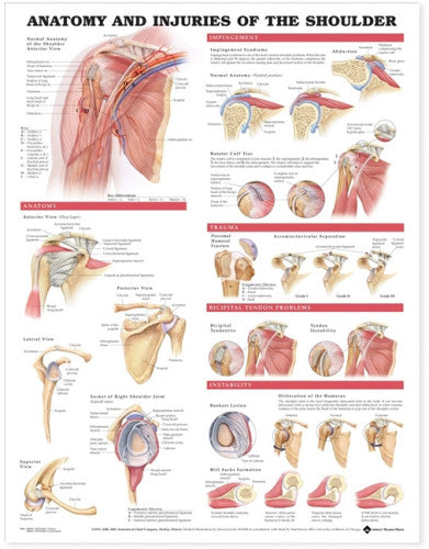
Shoulder anatomy chart. Shows muscles, ligaments and injuries.
The main image on this chart shows the bones, muscles, ligaments, veins, and arteries of the shoulder. The chart illustrates posterior, lateral, anterior, and superior views of the shoulder anatomy, as well as the socket of a normal shoulder joint. Images show impingement syndrome, rotator cuff tear, trauma (such as proximal humeral fracture and acromioclavicular separation), and bicipital tendon problems. The chart also illustrates instability such as anterior dislocation of the humerus, Bankart lesion, and Hill Sachs formation.
*Paper copy is available by special order.
 Therapist Recommends: Keep the anatomy right in front of you while you work.
Shipping Policy
In-stock items are shipped within 24 working hours otherwise, you will be notified. A shipping confirmation email with the tracking information will be sent to you when the order is shipped. We ship with Canada Post, Canpar, Cold Shot, Purolator, Fedex, and UPS along with several freight companies for pallet shipments. Additional charges may apply for remote locations, and shipping is limited to Canada.
Return Policy
Returns require prior approval and are subject to a 20% restocking fee, as well as shipping charges both ways. Items must be returned in original packaging and resalable condition, with a reason for the return. Returns are not accepted on opened oils, lotions, and creams, books, washed linens, videos, CDs, or used items. Special orders may not be returnable. Returns must be authorized within 14 days of receiving the product. For more information, please click on this link: Shipping and Returns Policy


